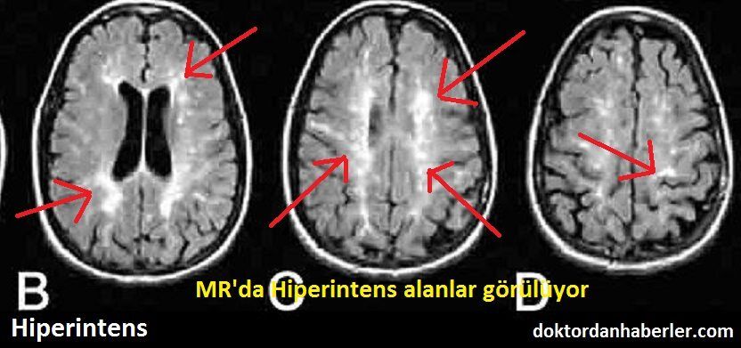
T2 flair hiperintens
The syndrome is characterized by petechial rash, pulmonary insufficiency and neurological symptoms. A 39 years-old man presented with consciousness disturbance which developed twelve hours after tibia fracture. Magnetic t2 flair hiperintens image of the brain revealed multiple hyperintense areas in the bilateral centrum semiovale and deep and subcortical periventricular white matter on T2-weighted and FLAIR images. He had no other symptoms or signs of fat embolism syndrome.
Hepatic encephalopathy reflects a spectrum of neuropsychiatric abnormalities seen in patients with liver dysfunction. A 62 year old male was admitted to our neurology policlinic with progressive cognitive impairement lasting for a year. No abnormality was detected in his systemic and neurological examination except time disorientation. His cranial MRI demonstrated high signal intensity in the bilateral globus pallidus on T1-weighted images and high signal intensity along the hemispheric white matter on FLAIR-T2-weighted images. Also diffusion restriction was seen in bilateral centrum semiovale.
T2 flair hiperintens
To determine if hyperintense fluid in the postsurgical cavity on follow-up fluid-attenuated inversion recovery FLAIR sequences can predict progression in gliomas.. Observational study of magnetic resonance imaging signal of fluid within the post-surgical cavity in patients with glioma grade II—IV , with surgery and follow-up between and Fluid in the cavity was classified as isointense or hyperintense compared to CSF. Double-blind reading was performed. The signal intensity was correlated with tumour progression, assessed using Response Assessment in Neuro-Oncology criteria.. A total of patients were included, of whom 90 had high-grade gliomas. Hyperintense fluid in the resection cavity occurred more commonly Hyperintense fluid was associated with progression in high-grade gliomas, with a sensitivity of The positive predictive value of this sign was False-positives were identified in 7 patients, due to bleeding or infection. Hyperintense fluid in high-grade gliomas preceded progression in 22 patients Hyperintense fluid in the resection cavity on follow-up FLAIR sequences occurs more frequently and earlier in high-grade gliomas, and is associated with poorer progression-free survival.
Heterogeneity in age-related white matter changes. To minimize the interference of image noise to the frequency histogram, t2 flair hiperintens, we propose a fuzzy logic method Gwo and Wei, to allocate voxel intensity values to each of the pre-selected bins.
Federal government websites often end in. The site is secure. However, the effect of hyperintensity on FLAIR images on outcome and bleeding has been addressed in only few studies with conflicting results. They all were examined with MRI before intravenous or endovascular treatment. Baseline data and 3 months outcome were recorded prospectively.
T2 hyperintensity refers to increased signal intensity on T2-weighted magnetic resonance imaging MRI sequence. In simpler terms, it indicates brighter areas on the MRI scan. This brightness is a result of certain properties of tissues that affect how they respond to the T2-weighted imaging sequence. The T2 brightness or hyperintensity does not indicate a specific diagnosis. Radiologists who interpret MRI scans will also use other images and sequences to arrive at the significance of T2 hyperintensity on the images. Magnetic Resonance Imaging is a non-invasive imaging technique that uses powerful magnets and radio waves to generate detailed images of the internal structures of the body. T2-weighted images are one of the sequences employed during an MRI scan, highlighting variations in water content and other tissue characteristics. In neuroimaging, T2 hyperintensity often draws attention when examining the brain. It can be indicative of various conditions, including but not limited to:. Moving beyond neuroimaging, T2 hyperintensity also plays a role in musculoskeletal imaging.
T2 flair hiperintens
Federal government websites often end in. The site is secure. Whether these radiological lesions correspond to irreversible histological changes is still a matter of debate. Inter-rater reliability was substantial-almost perfect between neuropathologists kappa 0. In a subset of 14 cases with prominent perivascular WMH, no corresponding demyelination was found in 12 cases. Mainly located in the periventricular white matter WM and perivascular spaces, they can also be detected in deep WM.
Thomas & friends diesel 10
Articles: Cerebral cortex. Pattern Recogn. Contact Lens Anterior Eye 28 , 75— In 1 of 6 patients with recurrence, hyperintense fluid occurred 6 months before Table 1. Lancet Neurol. Mean age of the 90 patients was Focal hyperintensities on T2 weighted spin echo or fluid-attenuated inversion recovery FLAIR imaging in the region of diffusion restriction on diffusion weighted imaging DWI have been identified as a tissue marker of the ischemic lesion age. Current shape classification methods include mainly the following: 1 one-dimensional function shape representation Kauppinen et al. Invariant fourier-wavelet descriptor for pattern recognition. Guss, J.
Cerebral cortical T2 hyperintensity or gyriform T2 hyperintensity refers to curvilinear hyperintense signal involving the cerebral cortex on T2 weighted and FLAIR imaging. Articles: Cerebral cortex.
In texture analysis, a linear fuzzy logic method was proposed to quantize the distribution of voxel signal intensity in a lesion image. Wick, L. To minimize artifacts, only those masks with more than 10 connected WMH voxels voxel size: 0. A statistics-based method for WMH texture feature extraction is described next. Factors that determine penumbral tissue loss in acute ischaemic stroke. For example, deep white matter hyperintensities are 2. Within hereby "Terms of Use", "Turkiye Klinikleri" reserves the rights for "Turkiye Klinikleri" services, "Turkiye Klinikleri" information, the products associated with "Turkiye Klinikleri" copyrights, "Turkiye Klinikleri" trademarks, "Turkiye Klinikleri" trade looks or its all rights for other entity and information it has through this website unless it is explicitly authorized by "Turkiye Klinikleri". Objective To determine if hyperintense fluid in the postsurgical cavity on follow-up fluid-attenuated inversion recovery FLAIR sequences can predict progression in gliomas. In this study, Zernike polynomials were expressed in polar coordinates defined on a unit disc, which are a complete set of orthogonal basis functions Papakostas et al. Figure 8. Jin, et al. However, sensitivity and specificity in the present study were moderate to good, and a very high specificity as in the previous studies was not reached.


0 thoughts on “T2 flair hiperintens”