Prostate pathology outlines
Maintenance between March 11 and 12 may cause some brief downtime. We apologize for any inconvenience!
Federal government websites often end in. The site is secure. This review article focuses on prostate carcinoma and underscores changes in the prostate chapter as well as those made across the entire series of the 5th edition of WHO Blue Books. Evolving and unsettled issues related to grading of intraductal carcinoma of the prostate and reporting of tertiary Gleason pattern, the definition and prognostic significance of cribriform growth pattern, and molecular pathology of prostate cancer will also be covered in this review. The publication of WHO Classification of Urinary and Male Genital Tumors 5th Edition marks another major milestone in the field of genitourinary GU pathology and is the culmination of scientific advancements in recent years built upon the 4th edition published in The new edition of this authoritative reference book provides a comprehensive update on tumor classification in the same modular fashion as the previous edition with the addition of several new sections for each disease entity, including cytology, diagnostic molecular pathology, essential and desirable diagnostic criteria, and staging. This review article highlights salient changes made to the prostate chapter as we have gained better understanding of the etiology, pathogenesis, and molecular pathology of prostate cancer.
Prostate pathology outlines
Maintenance between March 11 and 12 may cause some brief downtime. We apologize for any inconvenience! Epstein, M. Page views in 30, Cite this page: Samarska I, Epstein J. Accessed March 11th, Alpha methylacyl CoA racemase AMACR is a mitochondrial and peroxisomal enzyme, a amino acid protein essential in lipid metabolism, encoded by a bp sequence gene, located on chromosome 5p13 J Clin Pathol ; One of the most widely used markers of prostate carcinoma, because this protein is upregulated in prostate carcinoma and not found in benign prostate tissue J Clin Pathol ; , Am J Surg Pathol ;e6. Essential features. AMACR catalyzes the racemization of alpha methyl branched carboxylic coenzyme A thioesters and this enzyme is essential in the oxidation of bile acid intermediates and branched chain fatty acids J Clin Pathol ; Phytanic acid, present in red meat and dairy products, is one of the primary substrates of AMACR and has been found to be elevated in prostate adenocarcinoma Prostate ; AMACR is important in the pharmacological activation of ibuprofen and related drugs Bioorg Chem ; Clinical features.
Nonspecific granulomatous prostatitis. At this time, either method can be used, but pathologists should prostate pathology outlines which of the two is used for clarity and meaningful analyses in the future.
Maintenance between March 11 and 12 may cause some brief downtime. We apologize for any inconvenience! Page views in 6, Prostate specific antigen PSA. Accessed March 11th, Androgen regulated serine protease Encoded by kallikrein gene KLK3 , kallikrein related peptidase 3 located on chromosome 19 Endocr Rev ; Essential features.
Federal government websites often end in. The site is secure. This review article focuses on prostate carcinoma and underscores changes in the prostate chapter as well as those made across the entire series of the 5th edition of WHO Blue Books. Evolving and unsettled issues related to grading of intraductal carcinoma of the prostate and reporting of tertiary Gleason pattern, the definition and prognostic significance of cribriform growth pattern, and molecular pathology of prostate cancer will also be covered in this review. The publication of WHO Classification of Urinary and Male Genital Tumors 5th Edition marks another major milestone in the field of genitourinary GU pathology and is the culmination of scientific advancements in recent years built upon the 4th edition published in The new edition of this authoritative reference book provides a comprehensive update on tumor classification in the same modular fashion as the previous edition with the addition of several new sections for each disease entity, including cytology, diagnostic molecular pathology, essential and desirable diagnostic criteria, and staging. This review article highlights salient changes made to the prostate chapter as we have gained better understanding of the etiology, pathogenesis, and molecular pathology of prostate cancer. The following topics will be presented in detail: 1 changes in nomenclature and terminology, 2 prostatic ductal adenocarcinoma and prostatic intraepithelial neoplasia PIN -like adenocarcinoma, 3 intraductal proliferative lesions and reporting recommendations from the two major urological societies regarding intraductal carcinoma of the prostate, 4 cribriform growth pattern, 5 reporting of tertiary Gleason pattern, 6 treatment-related neuroendocrine prostatic carcinoma, and 7 molecular genetics. In addition to the updates specific to this chapter, the format of the contents had also been restructured across all volumes of the 5th edition series and that pertaining to the prostate chapter will be addressed first to provide an overview of how the new WHO Blue Book is organized. In alignment with the new format in the 5th edition series, less common but identical neoplasms from various sites in the GU system, i.
Prostate pathology outlines
Return to the tutorial menu. The male prostate gland is located below the bladder. The seminal vesicles are located posterior to the prostate. The urethra exits from the bladder and traverses the prostate before exiting to the penile urethra. The normal prostate is composed of glands and stroma. The glands are seen in cross section to be rounded to irregularly branching. These glands represent the terminal tubular portions of long tubuloalveolar glands that radiate from the urethra. The glands are lined by two cell layers: an outer low cuboidal layer and an inner layer of tall columnar mucin-secreting epithelium. These cells project inward as papillary projections.
Giants stadium seating chart
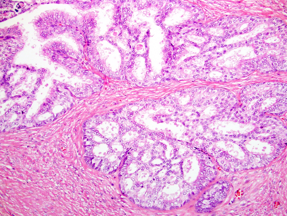
Robinson: The proportion of cases that including or excluding IDC-P would have changed the grade in these studies is very small and would not have any significant impact. The transition between both components is usually abrupt, and the concomitant PCa is often high-grade Fig. Historical and contemporary perspectives on cribriform morphology in prostate cancer. IHC stain for p63 demonstrates loss of basal cells in the cancer glands B, x and preserved basal cell layer in benign glands elsewhere in the same core not shown. At this time, either method can be used, but pathologists should specify which of the two is used for clarity and meaningful analyses in the future. Vascular invasion, extraprostatic extension, and seminal vesicle invasion may also be observed. For this reason, the diagnosis of cribriform HGPIN on needle biopsy should be avoided as this can lead to inappropriate patient management. Click here for information on linking to our website or using our content or images. Board review style question 2. Submit article Register Login.
Official websites use.
Answers B - D are incorrect because osteoclasts, syncytiotrophoblasts and sometimes neoplastic cells may be multinucleated, yet they would be found in different places bone tissue, placenta and tumors, respectively. Clinical features of neuroendocrine prostate cancer. Historical and contemporary perspectives on cribriform morphology in prostate cancer. Features diagnostic of cribriform glands. The proportion of cases that including or excluding IDC-P would have changed the grade in these studies is very small and would not have any significant impact. For localized PCa, several prognostic transcript signatures or genomic classifiers have been shown to improve risk stratification additional to clinicopathological features Molecular genetics of PCa initiation and progression, therapeutically relevant genomic alterations in metastatic PCa, and germline testing are succinctly discussed in several places in the prostate chapter. Jerasit Surintrspanont Ming Zhou. The squamous family comprises 3 tumor types: adenosquamous carcinoma, squamous cell carcinoma, and adenoid cystic basal cell carcinoma of the prostate. There is emerging evidence suggesting that IDC-P has two distinct biological pathways 36 despite the two being morphologically indistinguishable. Science ; Spread of adenocarcinoma within prostatic ducts and acini. Table II. Prostatic acinar adenocarcinoma. Neuroendocrine differentiation in prostate cancer: novel morphological insights and future therapeutic perspectives.


0 thoughts on “Prostate pathology outlines”