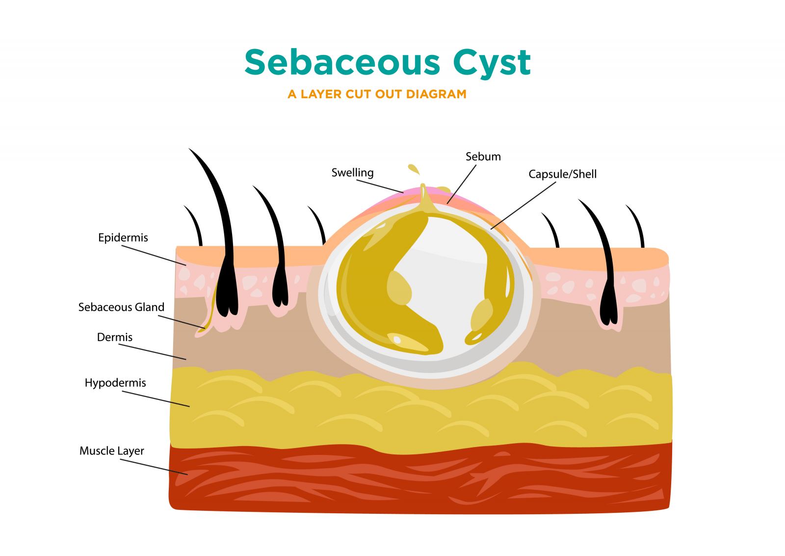
Pilar cyst diagram
DermNet provides Google Translate, a free machine translation service. Note that this may not provide an exact translation in all languages. Home arrow-right-small-blue Topics A—Z arrow-right-small-blue Trichilemmal cyst. Advisor to the content: Dr.
Pilar cysts, sometimes referred to as trichilemmal cysts or wens, are common growths that form from hair follicles; they are most often found on the scalp. Pilar cysts are smooth and mobile, meaning they can be moved slightly under the skin. They are filled with keratin a protein component found in hair, nails, and skin. They are usually painless but can be tender. There may be one or a few pilar cysts. Very rarely, pilar cysts can become cancerous.
Pilar cyst diagram
Pilar cysts grow around hair follicles and usually appear on the scalp. They are yellow or white and form small, round, or dome-shaped bumps. They grow slowly and may disappear on their own. In some cases, a doctor may remove them. A cyst is a small lump filled with fluid. They form under the skin. Cysts are very common and usually have no symptoms or side effects. There are three main types of cysts: epidermoid , meibomian chalazion , and pilar cysts. Many pilar cysts heal without treatment if a person is careful not to damage the skin. A surgeon is usually able to remove a cyst easily. However, sometimes even after removal, the cyst may reappear. A cyst will appear as a small, round, or dome-shaped bump. Some pilar cysts are yellow or white. Pilar cysts tend to be between 0. Because they grow very slowly, a person may not notice a pilar cyst until it reaches a certain size.
Acute inflammation after rupture is often pilar cyst diagram as a bacterial infection. Of all skin cysts, Pilar cysts are the most common cysts, mostly affect the skin of the scalp. Related Coverage.
Federal government websites often end in. Before sharing sensitive information, make sure you're on a federal government site. The site is secure. NCBI Bookshelf. Daifallah M. Al Aboud ; Siva Naga S.
Federal government websites often end in. Before sharing sensitive information, make sure you're on a federal government site. The site is secure. NCBI Bookshelf. Daifallah M. Al Aboud ; Siva Naga S.
Pilar cyst diagram
Pilar cysts tend to form on the scalp where keratin builds up in the lining of your hair follicles. They are typically harmless. Pilar cysts are flesh-colored bumps that can develop on the surface of the skin. You may be able to identify some of the characteristics of pilar cysts on your own, but you should still see your doctor for an official diagnosis. Keep reading to learn more about how these cysts present, whether they should be removed, and more. Pilar cysts grow within the surface of your skin. Although 90 percent of pilar cysts occur on the scalp, they can develop anywhere on the body.
Trovit
Pilar cysts may run in families. Pilar cysts occur most commonly in middle-aged women. The skin covering a pilar cyst is less fragile than that of an epidermoid cyst. Evaluation Diagnosis of pilar cysts is mainly clinical, based on signs and symptoms and additional investigations rarely need to be done. Kaya G, Saurat JH. Sequelae of trichilemmal cysts include inflammation, infection and malignant transformation which is rare. Trichilemmal cysts are derived from the outer root sheath of the hair follicle. Sign up to the newsletter. The site is secure. This activity illustrates the evaluation and treatment of pilar cysts and reviews the role of the interprofessional team in managing those with this condition.
Check out our latest pathology themed Wordle here! Updated every Monday. Page views in 56,
Postoperative and Rehabilitation Care After surgical removal of Pilar cyst, it is very important to taking care of the surgical site. Trichilemmal cysts are benign lesions, and they might transform into malignancy on rare occasions. The cyst shows very dense pink keratin on haematoxylin and eosin staining. A doctor can also recognize a pillar cyst easily under a microscope. Article Talk. The skin covering a pilar cyst is less fragile than that of an epidermoid cyst. Pilar Cyst. How to identify a pilar cyst? Enhancing Healthcare Team Outcomes Assessment of any patients with Pilar cyst begins with members of an interprofessional team of nurses and physicians taking a thorough history and careful physical examination which is usually enough to reach the diagnosis in most of the cases. Sequelae of trichilemmal cysts include inflammation, infection and malignant transformation which is rare. Of all skin cysts, Pilar cysts are the most common cysts, mostly affect the skin of the scalp.


Interesting theme, I will take part. Together we can come to a right answer.