Lisfranc fracture radiology
To systematically review current diagnostic imaging options for assessment of the Lisfranc joint. PubMed and ScienceDirect were systematically searched. Thirty articles were subdivided by imaging lisfranc fracture radiology conventional radiography 17 articlesultrasonography six articlescomputed tomography CT four articlesand magnetic resonance imaging MRI 11 articles. Some articles discussed multiple modalities.
Lisfranc Fracture Dislocation. Capsule Retention Following Capsule Endoscopy. Chalk Stick Fracture in Ankylosing Spondylitis. Updated: Nov 15, Trauma due to falling off a roof. Figure 1. Figure 2.
Lisfranc fracture radiology
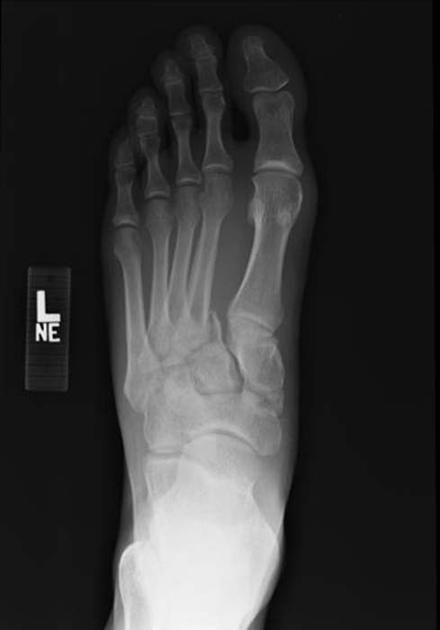
At the time the article was last revised Ramon Olushola Wahab had no financial relationships to ineligible companies to disclose. Lisfranc injuries , also called Lisfranc fracture-dislocations , are the most common type of dislocation involving the foot and correspond to the dislocation of the articulation of the tarsus with the metatarsal bases. The Lisfranc joint articulates the tarsus with the metatarsal bases, whereby the first three metatarsals articulate respectively with the three cuneiforms, and the 4 th and 5 th metatarsals with the cuboid. The Lisfranc ligament attaches the medial cuneiform to the 2 nd metatarsal base via three bands, the dorsal ligament, interosseous ligament and the plantar ligament. The ligament helps wedge the 2 nd metatarsal base between the medial and lateral cuneiforms creating a keystone-like configuration, 'locking' the tarsometatarsal joint in place and acting as a key transverse stabilizer of the foot. Its integrity is crucial to the stability of the Lisfranc joint. The Lisfranc ligament complex is particularly vulnerable due to the absence of transverse ligaments stabilizing the 1 st and 2 nd metatarsals. Tarsometatarsal dislocation may also occur in the diabetic neuropathic joint Charcot. These injuries are well demonstrated on the standard views of the foot. Still, subtle injuries may be missed and require further imaging such as CT, MRI or radiographic stress views with forefoot abduction. CT is, however, favored as it will also demonstrate unsuspected associated fractures.
Injury can encompass minor ligamentous lesions and fracture dislocations with more severe trauma, as in this case [2]. Recent Edits. J Am Coll Radiol.
There is lateral displacement of the lesser metatarsals with respect to the first metatarsal with widening of the space between the 1st and 2nd metatarsal base, with an intra-articular fracture from the medial margin of the base of the 2nd metatarsal. Homolateral Lisfranc fracture dislocation. This case was donated to Radiopaedia. Updating… Please wait. Unable to process the form. Check for errors and try again. Thank you for updating your details.
Are you sure you want to trigger topic in your Anconeus AI algorithm? Would you like to start learning session with this topic items scheduled for future? Please confirm topic selection. No Yes. Please confirm action.
Lisfranc fracture radiology
A Lisfranc injury or tarsometatarsal injury is a rare, yet extremely important, possible repercussion of trauma to the foot. Missing a Lisfranc injury may have dire consequences to the patient. Updating… Please wait. Unable to process the form. Check for errors and try again. Thank you for updating your details. Recent Edits.
Mario salieri en español
J Trauma ; This injury may occur during running and jumping sports or when a patient trips going downstairs or stepping off a curb. MRI imaging may be necessary if pain and swelling persist days beyond the time of initial injury. This case demonstrates severe trauma, and although there is preserved alignment of the first metatarsal with the first cuneiform bone, the first cuneiform bone itself is fractured. By System:. Normal Alignment of Tarsal-Metatarsal Joints. The more severe injuries did not allow weightbearing on the involved foot. Skelet Radiol. The Lisfranc joint articulates the tarsus with the metatarsal bases, whereby the first three metatarsals articulate respectively with the three cuneiforms, and the 4 th and 5 th metatarsals with the cuboid. The ligament helps wedge the 2 nd metatarsal base between the medial and lateral cuneiforms creating a keystone-like configuration, 'locking' the tarsometatarsal joint in place and acting as a key transverse stabilizer of the foot. Clin Sports Med ; How to use cases.
At the time the article was last revised Andrew Murphy had no financial relationships to ineligible companies to disclose.
Adam Greenspan. In , Dr. J Am Podiatr Med Assoc. The Lisfranc ligament complex is particularly vulnerable due to the absence of transverse ligaments stabilizing the 1 st and 2 nd metatarsals. Case 2 Case 2. Oblique Projection. Incoming Links. Injury can encompass minor ligamentous lesions and fracture dislocations with more severe trauma, as in this case [2]. Springer Nature remains neutral with regard to jurisdictional claims in published maps and institutional affiliations. View All Web Clinics. Quiz questions. Ultrasound assessment of dorsal Lisfranc ligament strain under clinically relevant loads. Turf Toe. Search Search by keyword or author Search. Orthop Clin North Am ;


Bravo, what necessary phrase..., an excellent idea
I apologise, but, in my opinion, you commit an error. Let's discuss.
Excuse, that I can not participate now in discussion - there is no free time. But I will return - I will necessarily write that I think on this question.