Fluorodeoxyglucose positron emission tomography
Yee C. Ung, Donna E. Maziak, Jessica A. Vanderveen, Christopher A.
Cancer Imaging volume 14 , Article number: 10 Cite this article. Metrics details. Histopathology or clinical-radiologic follow-up greater than 1 year was used as a reference. In all these clinical scenarios, adding ceCT resulted in a distinct benefit, by yielding a higher percentage of change in patient management. These data strongly underline the importance of strictly selecting patients for the combined exam.
Fluorodeoxyglucose positron emission tomography
Federal government websites often end in. Before sharing sensitive information, make sure you're on a federal government site. The site is secure. NCBI Bookshelf. Muhammad A. Ashraf ; Amandeep Goyal. Authors Muhammad A. Ashraf 1 ; Amandeep Goyal 2. Fludeoxyglucose F18 is a radioactive tracer that acts as a glucose analog and is used for diagnostic purposes in conjunction with positron-emitting tomography PET to localize the tissues with altered glucose metabolism. It does not have therapeutic use. Altered glucose metabolism has implications for malignancies, epilepsy, myocardial ischemia, inflammatory conditions, and Alzheimer disease. PET scan uses radiotracers injected into the patient before the scan to visualize the blood flow and metabolic and biochemical activities in diseased and healthy tissues. FDG is a glucose analog that tends to accumulate in the tissue with high glucose demand, like tumors and inflammatory cells. This activity describes the indications, mechanism of action in various conditions, administration, precautions in the particular patient population, and relevant information to maximize patient and staff safety while obtaining optimal images and analysis. Objectives: Identify the proper administration of fludeoxyglucose F
In general, lymph nodes less than 1. Br J Cancer 83—
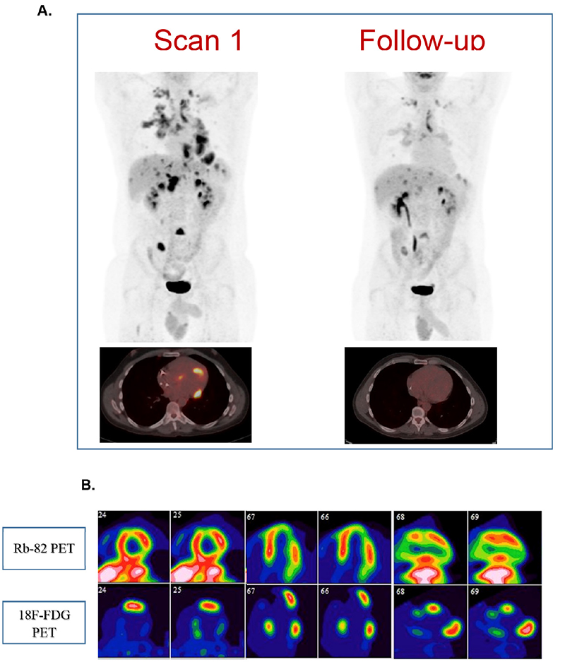
Initial studies of tuberculosis TB in macaques and humans using 18 F-FDG positron emission tomography PET imaging as a research tool suggest its usefulness in localising disease sites and as a clinical biomarker. Scanning was performed according to the EANM guidelines. One hundred and forty-seven patients with EPTB underwent 3 sequential scans. A progressive reduction over time of both the number of active sites and the uptake level SUVmax at these sites was seen. One died of brain tuberculoma.
Federal government websites often end in. The site is secure. Preview improvements coming to the PMC website in October Learn More or Try it out now. Rationale and objectives: Although low-dose computed tomography CT is a recommended modality for lung cancer screening in high-risk populations, the role of other modalities, such as [ 18 F]fluorodeoxyglucose-positron emission tomography PET , is unclear. We conducted a systematic review to describe the role of PET in lung cancer screening.
Fluorodeoxyglucose positron emission tomography
Positron emission tomography PET [1] is a functional imaging technique that uses radioactive substances known as radiotracers to visualize and measure changes in metabolic processes , and in other physiological activities including blood flow , regional chemical composition, and absorption. Different tracers are used for various imaging purposes, depending on the target process within the body. PET is a common imaging technique , a medical scintillography technique used in nuclear medicine.
Hypnosis gif
Fluorodeoxyglucose positron emission tomography in the evaluation of germ cell tumours at relapse. Implications of PET based molecular imaging on the current and future practice of medicine. Diabetes: Diabetes mellitus patients should have normal blood glucose for at least two days before administration. Disadvantages are that shot noise in the raw data is prominent in the reconstructed images, and areas of high tracer uptake tend to form streaks across the image. We present the current state of the art and speculate on the future applications of this technology including protocol development, newer imaging methods such as combined magnetic resonance and PET imaging and novel radiopharmaceuticals that can be used to further study tumor biology. Insulin causes the heart to increase GLUT4 receptors and promotes glucose uptake. The largest R 2 is last to enter. Due to these challenges, other cardiac diagnostic methods have been evaluated as potential tools to improve accuracy in the diagnostic evaluation of patients with IE. Discuss interprofessional team strategies for improving care coordination and communication to obtain suboptimal images while maintaining safety. Utility of positron emission tomography for the detection of disease in residual neck nodes after chemo radiotherapy in head and neck cancer. All imaging studies were interpreted by a team of experienced radiologists and nuclear medicine physicians well versed in myeloma diagnostics. Specificity remained high in all scenarios. Table 2 Distribution of lesions Full size table. Effectiveness of positron emission tomography in the preoperative assessment of patients with suspected non-small-cell lung cancer: the PLUS multicentre randomised trial.
Cancer Imaging volume 16 , Article number: 35 Cite this article.
Although coincidence scanners are no longer marketed, these devices continue to be used in some settings. Nomori a High rate of detection of unsuspected distant metastases by pet in apparent stage III non-small-cell lung cancer: implications for radical radiation therapy. Accuracy of positron emission tomography for diagnosis of pulmonary nodules and mass lesions: a meta-analysis. A further advantage of statistical image reconstruction techniques is that the physical effects that would need to be pre-corrected for when using an analytical reconstruction algorithm, such as scattered photons, random coincidences, attenuation and detector dead-time, can be incorporated into the likelihood model being used in the reconstruction, allowing for additional noise reduction. Sequential 18 F-fluorodeoxyglucose positron emission tomography 18 F-FDG PET scan findings in patients with extrapulmonary tuberculosis during the course of treatment—a prospective observational study. One of the factors most responsible for the acceptance of positron imaging was the development of radiopharmaceuticals. FDG-PET is used to detect the sites of infection and inflammation, particularly orthopedic infections related to osteomyelitis and implanted prostheses. Retrieved 11 December Please check for further notifications by email. Rights and permissions From twelve months after its original publication, this work is licensed under the Creative Commons Attribution-NonCommercial-Share Alike 3.


Excuse, I have removed this question
As that interestingly sounds
This theme is simply matchless :), very much it is pleasant to me)))