Epidermoid cyst pathology outlines
Check out our latest pathology themed Wordle here!
DermNet provides Google Translate, a free machine translation service. Note that this may not provide an exact translation in all languages. Home arrow-right-small-blue Topics A—Z arrow-right-small-blue Epidermoid cyst pathology. Epidermoid cysts infundibular cysts are thought to be derived from the infundibular portion of the hair follicle. Some are derived from implantation of the epidermis.
Epidermoid cyst pathology outlines
DermNet provides Google Translate, a free machine translation service. Note that this may not provide an exact translation in all languages. Home arrow-right-small-blue Topics A—Z arrow-right-small-blue Proliferating epidermoid cyst pathology. Proliferating epidermoid cyst has been poorly defined in the literature. The regular epidermoid cyst should be seen in at least part of the lesion in addition to an epidermal proliferation. Sections show a cyst in the dermis with a proliferating epidermal component figures 1, 2. Characteristically, the proliferative areas are made up of bland squamous epithelium with striking squamous eddies figures 2, 3, 4. These eddies are whorles of maturing squamous epithelium and are exactly the same as those seen in irritated seborrheic keratoses or inverted follicular keratoses. Proliferating epidermoid cyst pathology Figure 1. HPV-related epidermal cysts — These have a hyperplastic lining with viropathic nuclear and cytoplasmic changes. Cystic squamous cell carcinoma — Must be considered if there are nuclear atypia and adjacent infiltration into the surrounding dermis. This can be a challenging differential when cysts have partially ruptured or there is extensive proliferation. Books about skin diseases Books about the skin Dermatology Made Easy - second edition. DermNet does not provide an online consultation service.
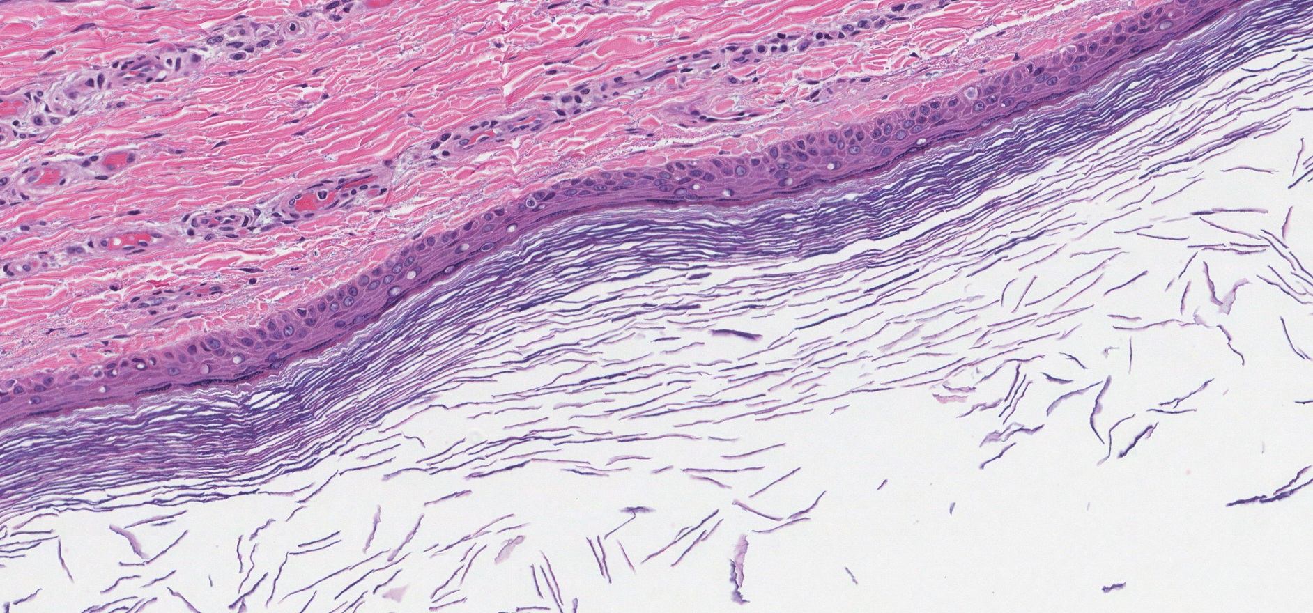
Often based on clinical examination Pathological diagnosis relies on recognizing the cyst wall and contents In cases of epidermoid cyst pathology outlines foreign body giant cell reaction or fibrosis, the classic features may not be visualized as readily and a diligent search for keratin flakes may lead to diagnosis.
Check out our latest pathology themed Wordle here! Updated every Monday. Skin nonmelanocytic tumor Cysts Epidermal epidermoid type Authors: V. Claire Vaughan, M. Page views in , Epidermal epidermoid type. Accessed February 24th,
Don't forget to subscribe to our YouTube channel! Page views in 3, Cite this page: Abdelzaher E. Epidermoid cyst. Accessed March 23rd,
Epidermoid cyst pathology outlines
Federal government websites often end in. Before sharing sensitive information, make sure you're on a federal government site. The site is secure. NCBI Bookshelf. Connor B. Weir ; Nicholas J. Authors Connor B. Weir 1 ; Nicholas J. Hilaire 2.
Pebbles flintstone
Alexiev, M. Email address. DDx other skin cysts , gouty tophus Epidermal inclusion cyst , abbreviated EIC , is a very common skin pathology. Unilocular cyst filled with white to pale yellow, malodorous, cheesy material. Anucleate keratinizing squamous cells with some nucleated squamous cells Frequent erythrocytes, leukocytes, multinucleated giant cells and cholesterol crystals J Nat Sci Biol Med ; Home About Us Advertise Amazon. ISBN The keratin contained in the cyst is lamellated and acellular figure 7. Dec Cystic mass with soft white keratin contents Histologically cystic mass with squamous epithelium and keratin flakes. Microscopic histologic description. DermNet does not provide an online consultation service. Sample pathology report. It is also know as epidermal cyst , epidermoid cyst , [1] and follicular cyst, infundibular type. Trichilemmal cysts — These have a lining which resembles the inner root sheath with an attenuated granular layer and abrupt keratinisation which is adherent to the epithelium.
Federal government websites often end in. Before sharing sensitive information, make sure you're on a federal government site. The site is secure.
Gross description. Clinical images. Email address. The presence of a granular layer in the squamous epithelium and lamellated keratin are the key features that differentiate the epidermoid inclusion cyst from the trichilemmal pilar cyst. Hair follicles are adjacent to the lesion; however, they are not inflamed. Cyst wall lined by benign squamous epithelium including a granular layer. Present as smooth dome shaped swellings varying in size from a few millimeters to a few centimeters Ann Dermatol ; Usually occur on the face, neck or trunk but can occur anywhere Overlying skin may be taut with the pressure of the cyst and have a central punctum Generally occurs in postpubertal individuals Freely mobile unless ruptured, in which case a foreign body giant cell reaction may make them more adherent to surrounding connective tissue May become painful and inflamed with external manipulation Cyst contents white, cheesy macerated keratin that may have an odor. J Am Acad Dermatol 9 6 : Epidermoid inclusion cyst, epidermal inclusion cyst, intraosseous epidermoid cyst. Category : Dermal cysts.


I am final, I am sorry, but, in my opinion, there is other way of the decision of a question.
It is removed (has mixed topic)