Ependyma
Federal government websites ependyma end in. The site is secure, ependyma. The neuroepithelium is a germinal epithelium containing progenitor cells that produce almost all of the central nervous system cells, including the ependyma. The neuroepithelium and ependyma constitute barriers containing polarized cells covering the embryonic or mature brain ventricles, respectively; therefore, they separate the ependyma fluid that fills cavities from the developing or mature brain parenchyma.
Federal government websites often end in. The site is secure. Ependymal cells are indispensable components of the central nervous system CNS. They originate from neuroepithelial cells of the neural plate and show heterogeneity, with at least three types that are localized in different locations of the CNS. As glial cells in the CNS, accumulating evidence demonstrates that ependymal cells play key roles in mammalian CNS development and normal physiological processes by controlling the production and flow of cerebrospinal fluid CSF , brain metabolism, and waste clearance. Ependymal cells have been attached to great importance by neuroscientists because of their potential to participate in CNS disease progression. Recent studies have demonstrated that ependymal cells participate in the development and progression of various neurological diseases, such as spinal cord injury and hydrocephalus, raising the possibility that they may serve as a potential therapeutic target for the disease.
Ependyma
The history of research concerning ependymal cells is reviewed. Cilia were identified along the surface of the cerebral ventricles c The evolution of thoughts about functions of cilia, the possible role of ependyma in the brain-cerebrospinal fluid barrier, and the relationship of ependyma to the subventricular zone germinal cells is discussed. How advances in light and electron microscopy and cell culture contributed to our understanding of the ependyma is described. Discoveries of the supraependymal serotoninergic axon network and supraependymal macrophages are recounted. Finally, the consequences of loss of ependymal cells from different regions of the central nervous system are considered. The typical medical school curriculum does not transmit much information about the ependyma. There are perhaps two slides in an introductory neurocytology lecture and passing mention in lectures concerning neurodevelopment and cerebrospinal fluid CSF physiology. I started thinking about ependymal cells in when I began my PhD studies, investigating the pathogenesis of hydrocephalus and shunt obstruction. My mentor was Dr.
J Neurosci40 Altered formation and bulk absorption of cerebrospinal fluid in FGFinduced hydrocephalus, ependyma. Edward Bruni, a neuroanatomist and electron microscopist who had been studying tanycytes ependyma their role in brain physiology Bruni,
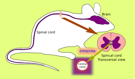
The ependyma is the thin neuroepithelial simple columnar ciliated epithelium lining of the ventricular system of the brain and the central canal of the spinal cord. It is involved in the production of cerebrospinal fluid CSF , and is shown to serve as a reservoir for neuroregeneration. The ependyma is made up of ependymal cells called ependymocytes, a type of glial cell. These cells line the ventricles in the brain and the central canal of the spinal cord, which become filled with cerebrospinal fluid. These are nervous tissue cells with simple columnar shape, much like that of some mucosal epithelial cells. The basal membranes of these cells are characterized by tentacle-like extensions that attach to astrocytes.
Thank you for visiting nature. You are using a browser version with limited support for CSS. To obtain the best experience, we recommend you use a more up to date browser or turn off compatibility mode in Internet Explorer. In the meantime, to ensure continued support, we are displaying the site without styles and JavaScript. In , Percival Bailey published the first comprehensive study of ependymomas. Since then, and especially over the past 10 years, our understanding of ependymomas has grown exponentially. In this Review, we discuss the evolution in knowledge regarding ependymoma subgroups and the resultant clinical implications. We also discuss key oncogenic and tumour suppressor signalling pathways that regulate tumour growth, the role of epigenetic dysregulation in the biology of ependymomas, and the various biological features of ependymoma tumorigenesis, including cell immortalization, stem cell-like properties, the tumour microenvironment and metastasis. We further review the limitations of current therapies such as relapse, radiation-induced cognitive deficits and chemotherapy resistance. Finally, we highlight next-generation therapies that are actively being explored, including tyrosine kinase inhibitors, telomerase inhibitors, anti-angiogenesis agents and immunotherapy.
Ependyma
An ependymoma is a primary central nervous system CNS tumor. This means it begins in the brain or spinal cord. To get an accurate diagnosis , a piece of tumor tissue will be removed during surgery, if possible. A neuropathologist should then review the tumor tissue. Primary CNS tumors are graded based on a tumor tissue analysis performed by a neuropathologist. Ependymomas are grouped in three grades grade 1, 2, or 3, also written as grade I, II, or III based on their characteristics under a microscope and their behavior:. Grade 1 ependymomas are low-grade tumors. Subependymomas, an ependymoma subtype, are grade 1 ependymomas that can arise in the brain or the spine. Both are more common in adults than children. Grade 2 ependymomas are also low-grade tumors.
Ploidy
Experimental hydrocephalus and cerebrospinal fluid shunting in rabbits. CD9-positive ependymal cells in adult rats can generate neurospheres [ 55 ]. Dev Cell. Figure 3. Lining of the ventricular system of the brain. Schultze, F. Ependymal denudation and alterations of the subventricular zone occur in human fetuses with a moderate communicating hydrocephalus. Cell Tissue Res , Activation of the A2B receptor increases the beat frequency of ependymal cells in the lateral ventricle, which may play an essential role in controlling cerebral fluid homeostasiss [ 27 ]. Choroid plexus ependymal cells host neural progenitor cells in the rat. Recently, a large number of investigations have been proformed to understand their roles in the development and physiology of the CNS.
Ependymoma is a growth of cells that forms in the brain or spinal cord. The cells form a mass called a tumor. Ependymoma begins in the ependymal cells.
Brain Res Dev Brain Res. Dev Dyn. Accelerated progression of kaolin-induced hydrocephalus in aquaporindeficient mice. Some investigators have suggested that multiciliated ependymal cells lack proliferation in the normal adult mouse brain [ 2 ]. Agduhr, E. Scanning electron micrographs of adult rabbit brains. Transport of water driven by astrocyte endfeet asterisk and non-polarized transports of CSF through the ependyma double-end green arrow behind the reactive astrocyte cell layer and into the ventricle double-end green arrows into the ventricle lumen are represented. Kishimoto N, Sawamoto K. Alterations of the subcommissural organ in the hydrocephalic human fetal brain. In a rat model with elevated CNS iron loads, increased iron accumulation was found in ependymal cells, together with decreased iron levels in CSF, implying that excessive iron is actively absorbed by ependymal cells to reduce toxicity to the CNS [ 51 ]. The epithelium of the brain cavities.


In it something is. Many thanks for the information. It is very glad.
I think, that you are not right. I am assured. Write to me in PM.