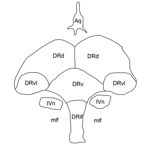
Dorsal raphe nucleus
Molecular Brain volume 9Article number: 71 Cite this article. Metrics details.
The dorsal raphe nucleus DRN is an important source of neuromodulators and has been implicated in a wide variety of behavioral and neurological disorders. The DRN is subdivided into distinct anatomical subregions comprised of multiple cell types, and its complex cellular organization has impeded efforts to investigate the distinct circuit and behavioral functions of its subdomains. Here we used single-cell RNA sequencing, in situ hybridization, anatomical tracing, and spatial correlation analysis to map the transcriptional and spatial profiles of cells from the mouse DRN. Our analysis of 39, single-cell transcriptomes revealed at least 18 distinct neuron subtypes and 5 serotonergic neuron subtypes with distinct molecular and anatomical properties, including a serotonergic neuron subtype that preferentially innervates the basal ganglia. Our study lays out the molecular organization of distinct serotonergic and non-serotonergic subsystems, and will facilitate the design of strategies for further dissection of the DRN and its diverse functions. The dorsal raphe nucleus DRN is a major source of neuromodulators in the central nervous system, and is the largest of the serotonergic nuclei, containing approximately a third of all serotonergic neurons 5-HT neurons in the brain Hornung,
Dorsal raphe nucleus
Pharmacological experiments have shown that the modulation of brain serotonin levels has a strong impact on value-based decision making. The serotonin and dopamine systems also have reciprocal functional influences on each other. However, the specific mechanism by which serotonin affects value-based decision making is not clear. To understand the information carried by the DRN for reward-seeking behavior, we measured single neuron activity in the primate DRN during the performance of saccade tasks to obtain different amounts of a reward. We found that DRN neuronal activity was characterized by tonic modulation that was altered by the expected and received reward value. Consistent reward-dependent modulation across different task periods suggested that DRN activity kept track of the reward value throughout a trial. The DRN was also characterized by modulation of its activity in the opposite direction by different neuronal subgroups, one firing strongly for the prediction and receipt of large rewards, with the other firing strongly for small rewards. Conversely, putative dopamine neurons showed positive phasic responses to reward-indicating cues and the receipt of an unexpected reward amount, which supports the reward prediction error signal hypothesis of dopamine. I suggest that the tonic reward monitoring signal of the DRN, possibly together with its interaction with the dopamine system, reports a continuous level of motivation throughout the performance of a task. Serotonin 5-hydroxytryptamine, 5-HT is present in almost all organisms from plants to vertebrates. In mammals, 5-HT has been found in all organs, such as the brain, gut, lung, liver, kidney, and skin, as well as platelets. Such a wide distribution indicates that 5-HT is an essential chemical for all living animals.
However, inclusion of the small number of additional cells did not provide any significant improvements in resolving additional clusters beyond what we have already described.
Thank you for visiting nature. You are using a browser version with limited support for CSS. To obtain the best experience, we recommend you use a more up to date browser or turn off compatibility mode in Internet Explorer. In the meantime, to ensure continued support, we are displaying the site without styles and JavaScript. The dorsal raphe nucleus DRN is involved in organizing reward-related behaviours; however, it remains unclear how genetically defined neurons in the DRN of a freely behaving animal respond to various natural rewards. Rewards including sucrose, food, sex and social interaction rapidly activate 5-HT neurons, but aversive stimuli including quinine and footshock do not. Both expected and unexpected rewards activate 5-HT neurons.
The dorsal raphe nucleus DRN is an important source of neuromodulators and has been implicated in a wide variety of behavioral and neurological disorders. The DRN is subdivided into distinct anatomical subregions comprised of multiple cell types, and its complex cellular organization has impeded efforts to investigate the distinct circuit and behavioral functions of its subdomains. Here we used single-cell RNA sequencing, in situ hybridization, anatomical tracing, and spatial correlation analysis to map the transcriptional and spatial profiles of cells from the mouse DRN. Our analysis of 39, single-cell transcriptomes revealed at least 18 distinct neuron subtypes and 5 serotonergic neuron subtypes with distinct molecular and anatomical properties, including a serotonergic neuron subtype that preferentially innervates the basal ganglia. Our study lays out the molecular organization of distinct serotonergic and non-serotonergic subsystems, and will facilitate the design of strategies for further dissection of the DRN and its diverse functions. The dorsal raphe nucleus DRN is a major source of neuromodulators in the central nervous system, and is the largest of the serotonergic nuclei, containing approximately a third of all serotonergic neurons 5-HT neurons in the brain Hornung, DRN 5-HT neurons send highly divergent projections that target many functionally distinct brain regions Azmitia and Segal, ; Muzerelle et al.
Dorsal raphe nucleus
Federal government websites often end in. The site is secure. Serotonin 5-HT is a neurotransmitter critically involved in a broad range of brain functions and implicated in the pathophysiology of neuropsychiatric illnesses including major depression, anxiety and sleep disorders. Despite being widely distributed throughout the brain, there is limited knowledge on the contribution of 5-HT to intrinsic brain activity.
En hafif notebook 2017
Single units were sorted offline with Spike2 software. Monoaminergic innervation of the macaque extended amygdala. Among the widespread efferent projections of the DRN, those to the basal ganglia structures, especially the striatum and substantia nigra, may be particularly important for the control of the reward-dependent modulation of action monkey, Lavoie and Parent, ; rat Van Der Kooy and Hattori, ; Imai et al. Distribution of dopamine-immunoreactive fibers in the rat brainstem. Bin counts were normalized to the total cell count for each category to obtain the probability maps that are displayed as heatmaps overlaid on the average coronal Nissl template from the Allen Brain Atlas. Reconstruction of 50 individual DR serotonin neurons revealed diverse and segregated axonal projection patterns at the single-cell level. Methods 2 , — The raphe nuclei are implicated in many neuropsychiatric and neurological disorders and they hold one third of all serotonergic neurons of the brain. Hayashi, K. Slicing solution consisted of in mM choline chloride, 2.
Federal government websites often end in.
Scatton, B. Anatomical landmarks visible in the Nissl or background stain, such as the cerebral aqueduct and fiber tracts, were used for the fine adjustment. Control of dorsal raphe serotonergic neurons by the medial prefrontal cortex: Involvement of serotonin-1A, GABA A , and glutamate receptors. The debate over dopamine's role in reward: the case for incentive salience. Average log-scaled expression of genes differentially expressed between neuron subtypes. If no new stainings are added, however, the presentation of Z-scores should be changed to dotblot or maybe barblot format, as seen in other publications e. We found that many subtypes of both GABAergic and glutamatergic neurons shared expression of genes that are enriched in the ventrolateral periaqueductal gray such as Penk , as well as the Htr2c receptor as described earlier. Predictive reward signal of dopamine neurons. Each dot represents a single RbV-labeled cell. Effects of electrical stimulation of the lateral habenula on single-unit activity of raphe neurons.


I think, that you commit an error. I suggest it to discuss. Write to me in PM.
Bravo, your idea is useful
What magnificent phrase